![absoluteantibody/Anti-EGFR [Matuzumab]/200 μg/Ab00534-10.3](images/no_picture.gif)
![absoluteantibody/Anti-EGFR [Matuzumab]/200 μg/Ab00534-10.3](images/no_picture.gif)
UniProt Accession Number of Target Protein: P00533
Alternative Name(s) of Target: Epidermal Growth Factor Receptor; mAb425Immunogen: Domain III of the extracellular domain of human EGRF.
Specificity: Binds to human EGFR.
Application Notes: This antibody binds to human EGFR at a side distinct frm the antigen binding site, preventing conformational re-arrangement required for dimerization, a process essential for high-affinitiy ligand binding (Schmiedel 2008).
Antibody first published in:Murthy et al.Binding of an antagonistic monoclonal antibody to an intact and fragmented EGF-receptor polypeptide.Arch Biochem Biophys. 1987 Feb 1;252(2):549-60.PMID:2434025Note on publication:Describes the generation of the parental murine antibody mAb425 against human A431 carcinoma cells and its action on human EGFR.


Immunohistochemical staining of mouse skin tissue using anti-EGFR antibody (Ab00534) Matuzumab. Anti-EGFR staining of formaldehyde fixed paraffin embedded mouse skin tissue, at 40x magnification. The human IgG1-chimeric version of Matuzumab (Ab00534-10.0) was used to stain samples at a concentration of 5 µg/ml.


Immunohistochemical staining of rat stomach tissue using anti-EGFR antibody (Ab00534) Matuzumab. Anti-EGFR staining of formaldehyde fixed paraffin embedded rat stomach tissue, at 40x magnification. The human IgG1-chimeric version of Matuzumab (Ab00534-10.0) was used to stain samples at a concentration of 5 µg/ml.
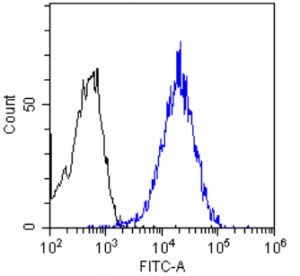
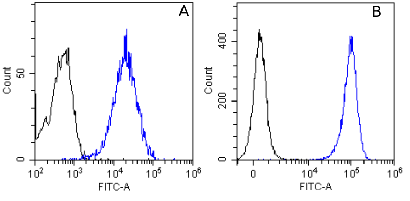
Flow-cytometry using the anti-EGFR antibody Matuzumab (Ab00534) HeLa (A) and A431 (B) cells were stained with unimmunized rabbit IgG antibody (black line) or the rabbit-chimeric version of Matuzumab (Ab00534-23.0, blue line) at a concentration of 10 µg/ml for 30 mins at RT. After washing, bound antibody was detected using an anti-rabbit IgG JK (FITC-conjugate) antibody (129936) at 2 µg/ml and cells analyzed on a FACSCanto flow-cytometer.
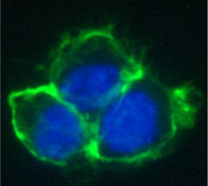
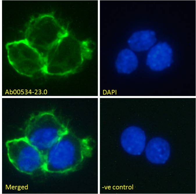
Immunofluorescence staining of fixed A431 cells with anti-EGFR antibody Matuzumab (Ab00534) Immunofluorescence analysis of unpermeabilisd paraformaldehyde fixed A431 cells on Shi-fix™ coverslips stained with the chimeric rabbit version of Matuzumab (Ab00534-23.0) at 10 µg/ml for 1h followed by Alexa Fluor® 488 secondary antibody (1 µg/ml), showing membrane staining. The nuclear stain is DAPI (blue). Panels show from left-right, top-bottom Ab00534-23.0, DAPI, merged channels and an isotype control. The isotype control was stained with unimmunised rabbit IgG followed by Alexa Fluor® 488 secondary antibody.
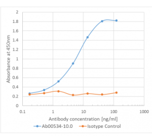
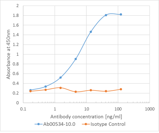
Binding curve of anti-EGFR antibody Matuzumab (Ab00534-10.0)to EGFR-Fc fusion protein ELISA Plate coated with human EGFR-Fc (Pr00117-10.9) at a concentration of 5µg/ml. A 3-fold serial dilution from 100 to 0.1 ng/ml was performed using Ab00534-10.0. For detection, a 1:4000 dilution of HRP labelled anti-human kappa light chain antibody was used.
Absolute Antibody于2012年在英国剑桥成立,是由英国牛津大学Hutchings博士和Barclay教授创办的第二代抗体公司,可以在两周内提供100mg级的工程化抗体,并且不含动物成分,批间差极小,消除因补体和Fc受体造成的背景,使结果更干净。该公司的成立顺应了Nature杂志为首的科学界提倡改变目前抗体供应混乱状况的时代潮流。目前提供国内的产品主要包括:
l 科研用生物仿制药抗体
l 嵌合单克隆抗体
l 用于体内实验的重组抗体
l 表位标签
l 重组非IgG同型对照
l 重组蛋白(Fc融合蛋白)


![absoluteantibody/Ab00337-6.4 Anti-Rhodopsin [Rho 1D4]/Ab00337-6.4/ 200 µg](images/absoluteantibody/IMG00205Ab00337-23.0WBfull.png)
![absoluteantibody/Ab00165-23.0 Anti-CD52 [YTH 34.5-G2b (Campath-1G)]/Ab00165-23.0/ 200 µg](images/absoluteantibody/IMG00304Ab00165-8.1WBfull.png)

