![absoluteantibody/Anti-Fas [R-125224]/50 μg/Ab00802-21.0](images/no_picture.gif)
![absoluteantibody/Anti-Fas [R-125224]/50 μg/Ab00802-21.0](images/no_picture.gif)
UniProt Accession Number of Target Protein: P25445
Alternative Name(s) of Target: h-HFE7A; Tumor necrosis factor receptor superfamily member 6; CD95; CD-95; Apo-1; Apo 1; Apo-1 antigen; Apoptosis-mediating surface antigen FAS; FASLG receptorImmunogen: R-125224 is generated by the humanization of the murine HFE7A anti-Fas antibody by grafting the CDR regions to the framework regions of the human 8E10 antibody and substituting key framework residues from the murine antibody into the 8E10 sequence. The original HFE7A was derived from a hybridoma cell line generated by the fusion of NS1 myeloma cells with splenocytes from Fas-deficient mice which had been immunized with partially purified recombinant human Fas-AIC2A chimera protein consisting of the extracellular region of human Fas antigen (aa -16 to 150) and the extracellular region of the murine IL-3 receptor AIC2 (aa 3-423). The HFE7A hybridoma was selected after screening by flow cytometry for the production of antibodies with the ability to bind to the WR19L12a transformed murine T cell lymphoma cell line expressing human Fas or the L5178YA1 cell line expressing murine Fas, but not to the parental WR19L or L5178Y cells.
Specificity: R-125224 binds to the extracellular portion of human Fas at an eptiope consisting of the sequence RTQNTKCRCK (aa 105-114) (pmid: 11754745). Fas is a type I membrane protein which belongs to the tumor necrosis factor (TNF) receptor/nerve growth factor (NGF) receptor superfamily. It is able to transduce apoptotic signals into the cell when bound by its ligand FasL (Fas ligand), which is primarily expressed in activated T lymphoid-myeloid lineage cells, in the eye, in reproductive organs and in some tumors. The Fas-FasL system is known to play an important role in maintaining the immune system as mice with Fas-defective lymphoproliferation (lpr) and FasL-defective generalized lymphoproliferative disease (gld) mutations develop massive lymphadenopathy and autoimmune diseases.
Application Notes: R-125224 shows the same binding affinity and the same ability to induce apoptosis in WR19L12a cells that express human Fas as the parental murine HFE7A antibody. R-125224 selectively induces apoptosis in type I activated lymphocytes but not in type II cells. R-125224 is able to induce apoptosis in the human lymphoid cell lines H9 and SKW6.4, as well as activated human lymphocytes, when cross-linked with anti-hIgG secondary antibodies. The antibody is unable to induce apoptosis in HPB-ALL cells, Jurkat cells or human hepatocytes. R-125224 has been used in vivo where it has been shown to greatly reduce the number of activated human human CD3+ Fas+ T cells in a SCID mouse model possessing a functional human immune system. Fas antigen tissue distribution in cynomolgus monkeys with collagen-induced arthritis at the arm joint (CIA monkeys) has been studied using [125I]-Labeled R-125224. High radioactivity in the bone marrow, thymus, lungs, liver, adrenals, spleen, ovaries, axillary lymph node and mesenteric lymph node compared to the radioactivity in the plasma was observed, which correlates with Fas expression. Fas can also be detected by R-125224 by ELISA.
Antibody first published in:Haruyama et alHumanization of the Mouse Anti-Fas Antibody HFE7A and Crystal Structure of the Humanized HFE7A Fab FragmentBiol. Pharm. Bull. 25(12) 1537—1545 (2002)PMID:12499636Note on publication:Describes the generation of 5 humanized versions of the mouse HFE7A antibody and their properties were studied and compared with the parental antibody. The crystal structure of R-125224 was determined.
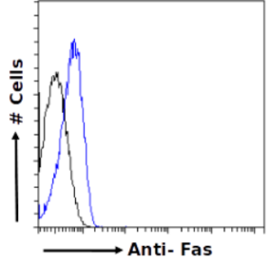
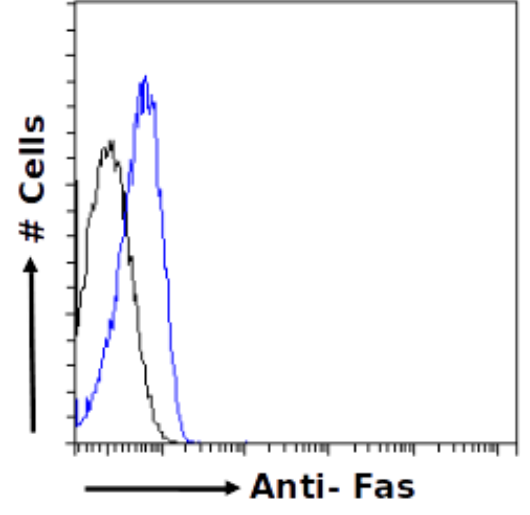
Flow-cytometry using the anti-Fas antibody R-125224 (Ab00802) Jurkat cells were fixed using 2% PFA, permeabilised using 0.5% Triton and stained with unimmunized rabbit IgG antibody (MOPC-21; isotype control, black line) or the rabbit IgG-chimeric version of R-125224 (Ab00802-23.0, blue line) at a dilution of 1:100 for 1h at RT. After washing, bound antibody was detected using a goat anti-rabbit IgG AlexaFluor® 488 antibody at a dilution of 1:1000 and cells analyzed using a FACSCanto flow-cytometer.
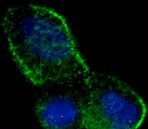
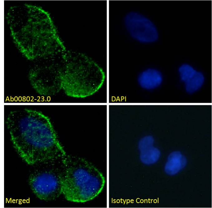
Immunofluorescence staining of fixed MCF7 cells with anti-Fas antibody R-125224 (Ab00802) Immunofluorescence analysis of paraformaldehyde fixed MCF7 cells permeabilized with 0.15% Triton and stained with the chimeric mouse IgG1 version of R-125224 (Ab00802-23.0) at 10 µg/ml for 1h followed by Alexa Fluor® 488 secondary antibody (2 µg/ml), showing membrane staining. The nuclear stain is DAPI (blue). Panels show from left-right, top-bottom Ab00802-23.0, DAPI, merged channels and an isotype control. The isotype control was stained with an anti-unknown specificity antibody (Ab178-23.0) followed by Alexa Fluor® 488 secondary antibody.
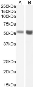
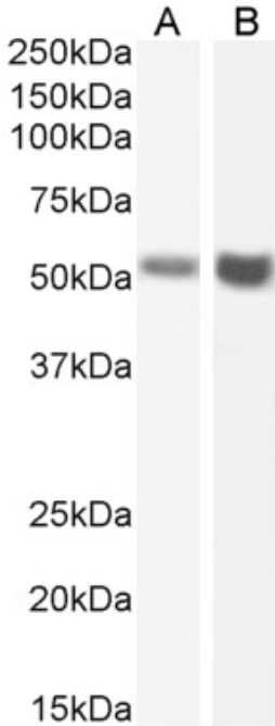
Western Blot using anti-Fas antibody R-125224 (Ab00802) Human testis (A) and human ovary (B) lysate samples (35µg protein in RIPA buffer) were resolved on a 10% SDS PAGE gel and blots probed with the chimeric rabbit IgG version of R-125224 (Ab00802-23.0) at 2 µg/ml before detection using an anti-rabbit secondary antibody. A primary incubation of 1h was used and protein was detected by chemiluminescence. The expected running size for unmodified Fas is 37.7kDa, but this protein is glycosylated at several positions leading to the observed running size.
Absolute Antibody于2012年在英国剑桥成立,是由英国牛津大学Hutchings博士和Barclay教授创办的第二代抗体公司,可以在两周内提供100mg级的工程化抗体,并且不含动物成分,批间差极小,消除因补体和Fc受体造成的背景,使结果更干净。该公司的成立顺应了Nature杂志为首的科学界提倡改变目前抗体供应混乱状况的时代潮流。目前提供国内的产品主要包括:
l 科研用生物仿制药抗体
l 嵌合单克隆抗体
l 用于体内实验的重组抗体
l 表位标签
l 重组非IgG同型对照
l 重组蛋白(Fc融合蛋白)


![absoluteantibody/Ab00337-6.4 Anti-Rhodopsin [Rho 1D4]/Ab00337-6.4/ 200 µg](images/absoluteantibody/IMG00205Ab00337-23.0WBfull.png)
![absoluteantibody/Ab00165-23.0 Anti-CD52 [YTH 34.5-G2b (Campath-1G)]/Ab00165-23.0/ 200 µg](images/absoluteantibody/IMG00304Ab00165-8.1WBfull.png)

